Ultrasound ultrasound refers to a technique used to examine internal organs in the abdomen and to guide a. 4 months ago, my first pet scan showed my breast tumor, one lymph node in my armpit and one lymph node in my chest.
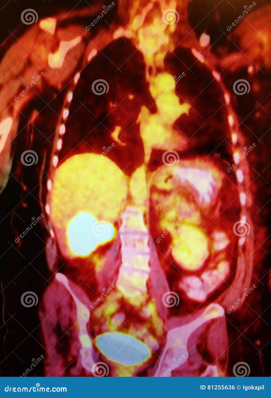
Patient with multiple metastatic lesions in liver & lung.
Pet scan images of liver cancer. Black and white ct scan of a cat pet on a black background. Pet/ct scans using axumin tracer, approved by fda last year and newly approved by medicare in some areas, are starting to be done at different locations. Since cancer cells intake more glucose than normal cells, injecting glucose into a vein and viewing the computerized image on a scan can reveal where the glucose concentrations.
[] showed that fdg uptake of hcc lesions correlates with the. The liver still shows metabolic activity (6.2 suv). These scans may help distinguish between benign and malignant tumors.
They may also be used to examine blood vessels in and around the liver. Should i request a liver biopsy ? Well, mom went from a relatively clear pet scan in may to a pet scan that shows several new places on her liver, more lymph node involvment in her lungs (i believe she said more spots there too) and three bone mets (lower back, right hip and left thigh.) the nurse practitioner was vague about exactly how many, i suppose she didn�t want to scare us.
A pet (positron emission tomography) scan combined with a ct scan is a specialised imaging test. For example, the ultrasound will travel through blood in the heart chamber, but the image will show when the echo of the heart is received. This test may help doctors determine whether the liver cancer has spread to areas such as the bones or lungs.
A positron emission tomography (or pet scan) is mainly used in cancer treatment, neurology, and cardiology. The pet/ct scan or pet/mri scan can help health professionals assess the degree of a disease’s impact. Patient with multiple metastatic lesions in liver & lung.
There is only one focus of increased activity, which is seen in the anterior aspect of the liver just cephalad to the gallbladder. Positron emission tomography (pet) scans of a patient with a rectum cancer and hepatic metastasis. The combined pet/ct scans provide images that pinpoint the location of abnormal metabolic activity within the body.
Procedure since any movement by the pet will make the images unreadable. After 13 rounds of chemo, my 2nd pet scan yesterday showed nothing in my breast or lymph nodes. Pet scans are special in that they can provide detailed images of nerves, arteries and tissues in addition to organs, bones and glands.
Its supposed to be more sensitive/specific than naf and other older scans, and comparable i think to choline or acetate (lots of studies and info on the web) and can. They are most often used for discovering and monitoring cancers of the soft tissues such as the brain, pelvis, liver, head, and neck. A pet (positron emission tomography) scan is a type of imaging test that uses radioactive glucose (radiotracer or radioactive tracer) to detect where cancer cells may be located in the body.
The combined scans have been shown to provide. One year later he developed liver metastasis, which was treated with radiofrequency ablation. A positron emission tomography (pet) scan can capture images that many other diagnostic tools fail to.
Liver imaging in patients with a history of known or suspected malignancy is important because the liver is a common site of metastatic spread, especially tumours from the colon, lung, pancreas and stomach, and in patients with chronic liver disease who are at risk for developing hepatocellular carcinoma. Similarly the sensitivity and specificity for metastases detection were 95% and 98% for pet/ct compared to 66% and 79% for pet alone. Ultrasound ultrasound refers to a technique used to examine internal organs in the abdomen and to guide a.
Today, most pet scans are performed on instruments that are combined pet and ct scanners. Liver cancer red rubber stamp over a white background. A liver pet scan may be used to inform a doctor of how well treatment is working for a diseased liver.
The ultrasound catches images when there is an echo or bouncing back of the organs. 4 months ago, my first pet scan showed my breast tumor, one lymph node in my armpit and one lymph node in my chest. Pet scans are much more sensitive than ct scans (see head and neck, lymph nodes, lung, more lung, liver , livermore liver, boneand bonefor some good examples.)
But the advantages of the superior clarity and resolution of ct scans can help identify cancer at earlier stages, thus increasing the likelihood of better outlook. The two scans provide more detailed and accurate information about where some cancers are in the body. A, fdg pet/ct scan done 10 months later shows recurrence with multiple hepatic and solitary pulmonary metastases.
Ct scans are often used with pet scans to diagnose cancer, examine heart muscle and detect brain abnormalities. Although cancer spreads silently in the body, pet can inspect all organs of the body for cancer in a single examination. Picture at the left is a pet scan showing colon cancer that has spread from the pelvis region to the liver.
Pet scan shows something in liver. Axial pet/ct image through the liver demonstrates.