Perhaps it may spot advanced lesions or masses but it doesn�t scan the entire area and can�t spot smaller representations of cancers. Four of eight patients in whom a loss of fatty hilum was seen in an axillary node on mri were found to have cancerous lymph nodes at the time of their breast surgery.

For example, a prostate biopsy is typically done with transrectal ultrasound and/or mri to help guide the biopsy.
Can you see cancer on an mri. Using mri, doctors can sometimes tell if a tumor is or isn�t cancer. Mri can also be used to look for signs that cancer may have metastasized (spread) from where it started to another part of the body. This means you won’t know whether or not you need to continue treatment.
Mris are used to determine if cancer has spread from your mouth to your neck. If you�re experiencing headache, fatigue, or memory problems, your physician will likely refer you for a brain tumor mri to confirm or rule it out. Doctors also use it to learn more about cancer after they find it, including:
A lymph node mri can be used to guide doctors or surgeons during a procedure, such as a biopsy. Mri scans have many other uses and can detect important conditions These pictures can show the difference between normal and diseased tissue.
An mri with contrast dye is the best way to see brain and spinal cord tumors. For example, it could be scar tissue left over from cancer killed off by your treatment. Rectal involvement is less common and can be seen as loss of posterior fat planes and direct tumor extension.
Mri creates pictures of soft tissue parts of the body that are sometimes hard to see using other imaging tests. From what i read (and i�m no doctor) an mri would pick up any future tumors of the type you had, so there still may be something that could be done to put your mind at ease about what you�re feeling. To plan cancer treatments, such as surgery or radiation therapy.
Mris can also show the size and extent of any cancer that has spread. Mri scans can show whether bladder cancer has spread to other tissues or to the lymph nodes. After cancer treatment, an mri is unable to determine whether remaining masses are cancerous:
Standard radiology specialty centers like ezra can assist patients needing mri with or without contrast imaging. In some cancers, such as cervix or bladder cancer, mri is better than ct at showing how deeply the tumour has grown into body tissues. Fifteen women had cancer in the nodes that required complete removal.
If radiologists don’t know which part of the body to be focusing on there will be plenty of false positives and false negatives. It’s typically safe, even if you’re pregnant. An mri scan uses radio waves and magnets to produce more detailed pictures of soft tissues.
The chances of a random mri picking up a cancer is pretty low because not all cancers are seen on mris. The world health organization says that 30 to 50% of cancers are preventable. Mri can also sometimes reveal other causes of pain in the tailbone region, such as cancer (tumor, malignancy), infection (abscess), pilonidal cyst (a painful lump.
The tool has produced encouraging results in a clinical. With companies like ezra, you can get screened without a physician’s advice and stay on top of your health. Four of eight patients in whom a loss of fatty hilum was seen in an axillary node on mri were found to have cancerous lymph nodes at the time of their breast surgery.
Because of this, physicians seek the help of an mri to look for issues in the male and female reproductive systems. The mri may show tissue that has cancer cells and tissue that does not have cancer cells. Unlike ct and positron emission tomography (pet) scans, mri scanners do not use ionizing radiation.
Perhaps it may spot advanced lesions or masses but it doesn�t scan the entire area and can�t spot smaller representations of cancers. Using mri, doctors can sometimes tell if a tumor is or isn’t cancer. Virtual mri or ct scan of the colon can detect polyp or mass but it can not diagnose cancer until a colonoscopy with biopsy is done.
Mri scans are used only for physical injuries reality: Many people do not know the difference between the two methods or why one might be selected over the other. An mri scan may be used if surgery is needed to remove a growth or lump.
The mri might show signs of cancer, but that cancer might not be active. Not all tumors are cancerous. Mri is very good at finding and pinpointing some cancers.
Mri is very good at finding and pinpointing some cancers. Part of this is due to early detection. Bladder involvement can be seen on mri as thickening of the posterior bladder wall and disruption of the hypointense bladder musculature or a mass within the bladder.
Ct (computed tomography) and mri (magnetic resonance imaging) are both used to diagnose and stage cancer. Which tests you might need will depend on the situation. It can be particularly useful for showing whether the tissue left behind after treatment is cancer or not.
Magnetic resonance imaging (mri) is a test that can be used to find a tumor in the body and to help find out whether a tumor is cancerous. Researchers have developed a new mri tool that can identify cases of ovarian cancer which are difficult to diagnose using standard methods. If you have an mri of the lower lumbar spine and nothing is noted in the colon area that it scans, this in.
It can help to show whether breast cancer has spread. If you are found to have prostate cancer, you might need imaging tests of other parts of your body to look for possible cancer spread. The size and location of the tumor.
An mri scan can show changes in the body by using magnetism and radio waves to create pictures. Radiation is never used under any circumstances and your mri scan will not cause cancer. For example, a prostate biopsy is typically done with transrectal ultrasound and/or mri to help guide the biopsy.
As others have said, i would look for another doctor who will take your concerns seriously and tell you what can be done instead of what can�t. Doctors use mris to take pictures of your spinal column, brain, chest, abdomen and breast. A research group at yale university school of medicine wondered, “if a man has a multiparametric mri (mpmri) of the prostate and it doesn’t show significant prostate cancer (pca), what are the chances that it’s wrong?” this is an excellent question, given that studies have shown that mpmri done by experts has roughly 98% accuracy in detecting significant cancer.
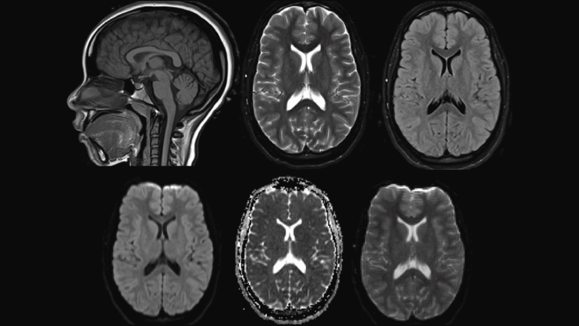
False positives give way to more unnecessary investigations because now there’s something you get to worry about. An mri with contrast dye is the best way to see brain and spinal cord tumors. Can you see a brain tumor on an mri scan?
By comparison, only 11 out of 48 patients, or 23 percent, with all fatty hilum in place had cancer. A lymph node mri scan may be done to check for certain cancers or other illness. To improve the quality of the images it’s sometimes necessary to administer an intravenous dye.